

Translational Science Benefits


Summary
One in 18 people (5 percent of the U.S. population) experience cardiac arrhythmia, a problem with the rate or rhythm of the heartbeat.1 Arrhythmias can lead to complications such as stroke, fainting and death. Treatments include medicine, lifestyle changes, and a procedure called a catheter ablation. Catheter ablation uses radiofrequency heat energy to destroy a small area of heart tissue that is causing rapid and irregular heartbeats.2 Typically, the physician has to mentally combine two 2D images of the heart displayed on different monitors to decide whether to remove tissue. This can cause a lot of mental fatigue.3
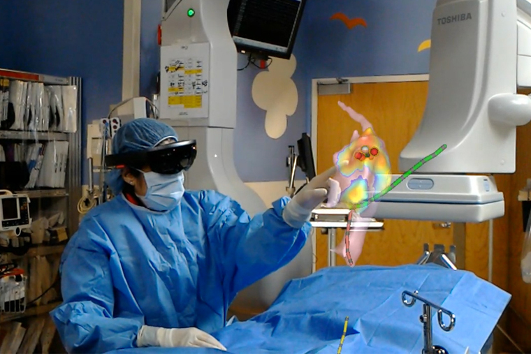
Husband and wife team Dr. Jonathan Silva and Dr. Jennifer Silva, MD, at Washington University in St. Louis, created the Enhanced Electrophysiology Visualization and Interaction System (ĒLVIS) to help physicians better visualize the interior of the heart during ablation procedures. ĒLVIS combines augmented reality (AR) software with Microsoft’s HoloLens headset to convert commercially available electroanatomic and catheter data into a 3D holographic image of the patient’s heart with real-time catheter locations. The physician uses their gaze and gestures to control the headset—optimizing their visualization and improving their accuracy—all while keeping their hands sterile for the procedure. And unlike virtual reality, the physician can still view their environment while the image “floats” over the patient.4
Significance
Cardiac ablation procedures require the physician to ablate very small areas of heart tissue (typically about 6 mm in size) while not damaging the rest of the heart muscle.5 The more intuitive 3D ĒLVIS display increases physician accuracy at removing tissue in the target area when compared to traditional 2D visualization, which is likely to improve patient outcomes and decrease the need for repeat procedures.5 ĒLVIS can also be used by multiple physicians using multiple headsets, improving coordination and supporting training.
Benefits
Demonstrated benefits are those that have been observed and are verifiable.
Potential benefits are those logically expected with moderate to high confidence.
Clinical & medical benefits
Developed CommandEP augmented reality (AR) software that creates a 3D digital image of the interior of the patient’s heart and can be displayed using Microsoft’s HoloLens headset. demonstrated.
Improved accuracy of cardiac ablation procedures by providing a real-time, 3D holographic image of the patient’s heart to help physician analyze heart tissue. demonstrated.
Community & public health benefits
Economic benefits
Founded SentiAR, a start-up company to market and sell ĒLVIS throughout the U.S. demonstrated.
Patented the ĒLVIS system and method for creating a 3D holographic image of the patient’s heart (U.S. Patent No: US10258426B2, April 2019.)6 demonstrated.
Reduced cardiac ablation procedure time by 30 minutes, freeing up hospital operating rooms for an additional 1-2 procedures per day, and may also reduce the need for the mapping technician role, generating additional cost savings for the hospital.7 potential.
Decreased need for repeat cardiac ablation procedures would save insurers an estimated $1,224 per procedure.7 potential.
Decreased need for repeat cardiac ablation procedures would save roughly $370 million per year, with the potential for savings to increase as the population ages and more people need cardiac ablation procedures.7 potential.
This research has clinical, community, and economic implications. The framework for these implications was derived from the Translational Science Benefits Model created by the Institute of Clinical & Translational Sciences at Washington University in St. Louis.8
Clinical
ĒLVIS combines an augmented reality (AR) platform with wearable headset technology to improve the accuracy of therapeutic cardiac ablation procedures to treat irregular heart rhythms. The proprietary software technology transforms electroanatomic and real-time catheter location information into a 3D holographic image of the heart that can be manipulated by physicians through their gaze and gestures without damaging the sterile field. Tests of the ĒLVIS display in humans found adequate performance, image quality, and usability of the software for ablation procedures.9
Community
ĒLVIS improves health care quality by enhancing the ability of physicians to visualize damaged cardiac tissue during ablation procedures, decreasing mental fatigue, and improving accuracy. ĒLVIS can significantly reduce the number of lesions that remain outside the target area from 36% in a typical ablation to just 4%.5 This improved accuracy is likely to reduce the need for repeat procedures. Patients with repeat ablations are more likely to visit the emergency department and require subsequent hospitalization.10
Economic
Drs. Jonathan and Jennifer Silva co-founded SentiAR, a startup company to develop and manufacture an augmented reality (AR) platform to transform patient and clinician experiences in interventional procedures. To develop the platform, the doctors secured a patent for the system and method used to integrate the AR data with the HoloLens headset. In September 2020, SentiAR received FDA clearance to market and distribute the platform to hospitals. By reducing the average procedure length and need for specialized the staff, ĒLVIS has the potential to be cost-effective for hospitals. By reducing the need for repeat procedures, the system could save insurers an estimated $1,224 per procedure, for a savings of $370 million annually.7
- Scripps Health. Top 10 Things You Should Know About Heart Rhythm; 2015. Accessed November 9, 2020.
- Ablation for Arrhythmias. American Heart Association. Published September 30, 2016. Accessed November 9, 2020.
- Early R. How an invention gets out of the lab and into the world. The Source. Published June 15, 2020. Accessed November 9, 2020.
- Miller B. WashU-developed holograms help physicians during cardiac procedure. The Source. Published July 20, 2020. Accessed November 9, 2020.
- Avari Silva JN, Southworth MK, Blume WM, et al. First-in-human use of a mixed reality display during cardiac ablation procedures. JACC Clin Electrophysiol. 2020;6(8):1023-1025. doi:10.1016/j.jacep.2020.04.036
- Silva J, Silva J. System and method for virtual reality data integration and visualization for 3D imaging and instrument position data. Published online April 16, 2019. Accessed November 9, 2020.
- V Capital. SENTIAR: Wearable Command Center in the Operating Room. Accessed September 3, 2020.
- Institute of Clinical & Translational Sciences at Washington University in St. Louis. Translational Science Benefits Model website. Published February 1, 2019. Accessed October 21, 2020.
- Southworth MK, Silva JNA, Blume WM, Hare GFV, Dalal AS, Silva JR. Performance evaluation of mixed reality display for guidance during transcatheter cardiac mapping and ablation. IEEE J Transl Eng Health Med. 2020;8:1-10. doi:10.1109/JTEHM.2020.3007031
- Mansour M, Karst E, Heist EK, et al. The impact of first procedure success rate on the economics of atrial fibrillation ablation. JACC Clin Electrophysiol. 2017;3(2):129-138. doi:10.1016/j.jacep.2016.06.002